In vitro studies
Prebiotics are used as substrates
In this research, four different oligosaccharides were utilized as prebiotics. The oligosaccharides included mannan oligosaccharide (Bio-Mos®), oligofructose (with an average degree of polymerization of 4) (Orafti®), galactooligosaccharides (GOS) (with an average degree of polymerization of 6), by Friesland Foods Domo of Holland, and xylooligosaccharides (with an average degree of polymerization of 3), by Shandong Longlive Bio-Technology of China.Prebiotics were kept in the recommended conditions by the manufacturing companies until the experiment began.
Probiotic strain
In this experiment, E. faecium (DSM 3530) was employed as the bacterium. The bacteria were identified using two primary tests, namely Gram and catalase staining tests.
Growth curve of E. faecium bacteria
To identify the bacterial growth pattern of E. faecium bacteria, the growth rate of this bacterium was investigated at different times in 30 h. Ninety cc of MRS broth culture medium was prepared and transferred to 30 test tubes (10 test tubes for 10 different times, each with 3 repetitions, and 3 cc samples in each test tube). The environment’s pH was adjusted to 5.8 ± 1 using hydrochloric acid and 1 normal NaOH. The culture medium temperature was maintained at 41 °C9 To create anaerobic conditions, the test tubes were covered with one cc of sterile liquid paraffin10. After autoclaving and sterilization, the media were inoculated with 1.5 × 10⁸ colony-forming units of E. faecium bacteria, previously cultured on MRS agar. To ensure the anaerobic conditions of the culture environment, the tubes were covered with cotton and aluminum foil after inoculation of bacteria. Then, the test tubes were transferred to a shaker incubator at 41 °C. In order to determine the concentration of bacteria, the test tubes were removed from the incubator at regular intervals after inoculation (every 3 h). Under sterile conditions, 1 cc samples were taken from them and transferred to glass cuvettes. Then, the optical absorption of each cuvette at a wavelength of 600 nm was read and recorded using a spectrophotometer (BRITE Technologies, Canada, Model BT 600)11.
Investigating the ability of E. faecium bacteria to consume prebiotics as a carbon source
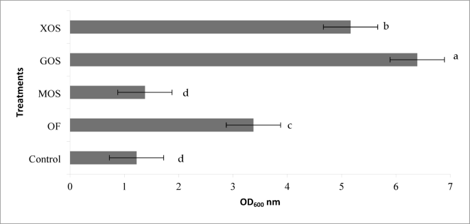
The study focused on the synbiotic impacts of E. faecium probiotic bacteria on 4 different polymerized prebiotics, namely mananoligosaccharide, oligofructose, GOS, and xyloligosaccharide, and their comparison to their corresponding control strains. Each prebiotic was considered a treatment, and three repetitions were used for each treatment. The pH, temperature, and anaerobic conditions were adjusted as described after the media was prepared. To prevent the reaction of prebiotics with the components of the environment during autoclaving, prebiotics were added to the environment separately and in the form of a 10% aqueous solution under sterile conditions. The treatment was supplemented with the same amount of distilled water without any probiotics. To inoculate the medium with prebiotics, a fresh culture of E. faecium bacteria was prepared 24 h before the experiment and diluted at 2.5 × 10⁸. After completing the preparation of the experimental treatments in the tubes, the medium was inoculated with E. faecium bacteria at the rate of 1% and with a dilution equal to 1.5 × 10⁸. To determine the growth rate of E. faecium bacteria in each of the treatments and to determine optical density, the test tubes were removed from the incubator at regular intervals after inoculation (every 3 h). Under sterile conditions, a 1 cc sample was taken from them. After removing it, it was transferred to glass cuvettes where it was read and recorded by a spectrophotometer at a wavelength of 600 nm11.
Determining the pH of the environment
The pH level of different treatments was investigated to determine and compare the acidity changes caused by the fermentation of different prebiotics by E. faecium bacteria in the culture medium. For this purpose, after measuring the optical absorption of the samples, the pH of different environments was read and recorded by a digital pH meter.
Number of live E. faecium bacteria in the exponential growth phase
To determine and compare the number of live bacteria in the culture medium containing different prebiotics, the counting of bacteria in the exponential growth phase was investigated. For this purpose, different environments were removed from the incubator 12 h after incubation, and 1 cc sample was taken from them under sterile conditions. After preparing the dilution series of the desired sample, the amount of 0.1 cc of the desired sample was spread on MRS solid culture medium. Finally, after 12 to 24 h of incubation at 41 °C, the number of bacteria was counted12.
In vivo studies
Birds, housing, and management
The animal trial was conducted in accordance with the guidelines was approved the Animal Care and Use Review Committee at Gorgan University of Agricultural Sciences and Natural Resources in Gorgan, Iran (Approval No. AK1390M/32/56). All protocols adhered to the ARRIVE guidelines for reporting animal research ( The mechanical cervical dislocation method was used for euthanasia, utilizing the Koechner Euthanizing Device, as recommended by the American Veterinary Medical Association. The required number of 400 broiler chickens of the Cobb 500 breed was procured from Tirgan Bandar’s mother hen company. At the time of egg production, the mother hen flock was in the first production cycle and at the age of 25 weeks, and the average initial weight of the chicks was 35 g, according to the hatching claim. The intended chickens were randomly assigned to 4 experimental treatments, each of which included five repetitions, and each repetition included 20 pieces of one-day-old chickens of two sexes. A control ration was prepared without using any growth-promoting antibiotic and anti-coccidiosis drug and based on the recommended requirements of the Cobb 500 breed. The desired additives were added to the control diet to prepare other treatments at suitable levels as follows. Experimental treatments were applied from the first day until 42 days. The combination of experimental treatments was as follows:
-
1.
Control;
-
2.
Control + prebiotic GOS (500 g per ton of feed).
-
3.
Control + E. faecium probiotic (500 g per ton of feed).
-
4.
Control + selected synbiotic.
Management
Before the arrival of chickens, the temperature was raised to 34 ℃. This temperature decreased by 3 ℃ every week until temperature reached 18 °C, and this temperature remained constant until the end. After entering, experimental chickens were nested randomly inside plastic cages (each pen was 220 × 170 cm2). At the beginning of the arrival of the chickens, water containing sugar and vitamin supplements was provided to them. The chicks had ad libitum to food and water from the first day. In the first week of rearing, water, and feed were provided to the chickens manually and in special floor trays for one-day-old chicks. From the second week to the end of the rearing period, the chickens were fed using hanging feeders and waterers whose height from the ground level was changed according to the age of the chickens. The lighting system was also adjusted based on Cobb 500 recommendations.
Diet preparation
Before preparing experimental rations, the amounts of nutrients in corn and soybeans, which comprised most of the experimental rations, were determined by the NIR device in the laboratory of Evonik Iran. Then, according to the amount of nutrients obtained and based on the recommended requirements of the Cobb 500 breed (2003), a basic diet for three initial periods (1 to 10 days old), grower (11 to 22 days old), and finisher (23 to 42 days) prepared and tested additives were added to it based on their degree of purity. To prepare the desired rations in each period, the values of the basic rations were first weighed for all treatments. Then, the desired amounts were mixed by a 500 kg horizontal mixer for 10 min. After mixing the rations, the amounts required for each experimental treatment were prepared then additives were added manually to the basic rations in several steps. Finally, the desired rations were pelleted by a pellet press machine under a controlled conditioner temperature of 70 ± 2 °C, and after cooling and bagging, they were made available to the birds. It should be noted that 2, 3, and 4 dyes were used to prepare the initial, grower, and finisher diet, respectively. Diet components and nutrient composition of experimental diets used are listed in Table 1.
Growth performance
Chickens of different experimental groups were weighed weekly using a digital scale. The body weight increase of birds was also calculated by subtracting the body weight obtained from the initial weight or the weight of the last week of chickens. It should be mentioned that the chickens were starved before weighing and because the digestive system was empty. The feed consumption of experimental units was determined weekly based on the difference between the amount of feed given at the beginning and the amount of feed remaining at the end of the week. The feed conversion ratio of experimental units was also calculated every week by dividing the amount of feed consumed by the experimental unit by the weight gain of the same experimental unit.
Morphology of the small intestine
To determine the effects of using the tested additives on the morphological characteristics of the small intestine, at the age of 27 and 42 days, five birds from each experimental treatment with a body weight close to the average of each experimental unit were selected. After slaughtering these birds, 2 cm of the middle part of their jejunum was separated, and the food contents in them were slowly removed using geyser. After the initial fixation of the tissues in 4% formalin buffer, the tissues were removed from the solution. After making a longitudinal cut with a surgical blade, they were again kept in formalin until staining and future investigations. Then, five-micron-thick slices were prepared from the jejunum and placed on glass slides. The samples were deparaffinized by xylan solution and dehydrated in graded alcohol solutions. Finally, the samples were stained with hematoxylin and eosin, and the height of the villi, the thickness of the villi, and the depth of the crypts were determined using a light microscope equipped with a camera and analyzed by Image Analysis software13.
Counting the bacterial population and pH of the gastrointestinal tract
One chicken piece was randomly selected from each experimental unit to determine the effects of experimental treatments on the pH and the number of microbial populations in the digestive system at the age of 27 days. After slaughter, it was taken to the laboratory. After disinfecting the abdominal surface of the carcass, the samples obtained from the contents of the ileum and cecum were homogenized with 0.9% saline solution at a ratio of 1:9. After diluting the obtained samples, Lactobacillus, coliform, and Escherichia coli were cultured in their specific culture media. The number of colonies was counted depending on the growth rate of the desired bacteria after 24 to 72 h of incubation at 37 °C. The counting results were multiplied by the dilution ratio and converted into logarithmic data. The pH of the ileum and cecum of slaughtered birds was also read and recorded using a digital pH meter.
Determination of antibody titer against Newcastle virus
To determine and compare the antibody titer against Newcastle virus in different experimental treatments, on the 23d day of the experiment, one chicken was selected from each experimental unit whose weight was as close as possible to the average weight of that experimental unit, and 2 ml of blood was collected through the subclavian vein. Immediately after blood collection, the intended chickens were marked with a spray, and 10 days later, blood collection was done again from the same chickens. After transfer to the laboratory, the antibody titer against Newcastle was determined by the samples’ agglutination inhibition (HI) method (European Pharmacopoeia, 2004). In short, the obtained blood samples were centrifuged for 10 min at 15,000 rpm to prepare serum and blood serum was extracted. 25 µl of serum from blood samples were added to 25 µl of phosphate-buffered saline solution of the first dilution of a 96-well plate, and dilution series was prepared with PBS. After dilution, 25 µl of the antigen solution, which was determined using the HA test, or the amount of 4 units of HA was controlled using the back titration test, was added to all the wells except the first well. The desired plate was incubated and placed on a mechanical stirrer for 30 min at room temperature. Then, 25 µl of 1% red blood cell solution were washed three times with PBS solution and added to all the wells, and the desired plate was again placed on a mechanical stirrer for 30 min at room temperature. Finally, the amount of antibody titer was read and recorded by the log2 dilution of the first well in which the patch was formed.
Statistical analysis
This experiment was conducted as a completely randomized design with four treatments and five repetitions. The one-way analysis of variance test of the SAS software package (2003) was used to analyze the data obtained from this experimental stage. A comparison of means was also done with Duncan’s multi-range test at a significance level of 5%.
link

